
Figures & data
Figure 1 On admission to the hospital, X-ray and MRI images were taken of the left knee joint. (A and B) An X-ray of the distal femur and the proximal tibia revealed patches of osteopenia (blue circle). (C–E) There was a synovial infection in the knee joint, a localized full-thickness defect in the patellar cartilage (red arrow), and a diffuse patchy signal in the bone marrow on T2-weighted imaging (red circle).
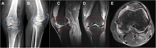
Figure 2 Brucella melitensis was detected by real-time PCR, real-time PCR showed that the DNA content of Brucella melitensis (Solid red line) increased in 32 cycles.
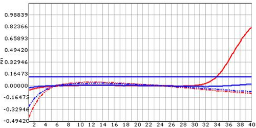
Figure 3 Pathological staining of knee joint effusion and inflammatory tissue. (A) Inflammatory cell infiltration was seen by HE staining. (B) Acid-fast staining was negative.
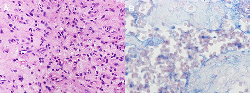
Figure 4 After five years, X-rays and MRI scans of the left knee joint were taken. (A and B) The X-rays showed that the distal femur and proximal tibia had resolved from the previous abnormal X-ray appearances. (C–E) It was also noted that the MRI changes were largely resolved.
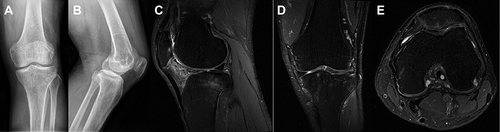
Table 1 Literature Review of Knee Joint Tuberculosis with Mixed Infections