Figures & data
Figure 1 Painful erythematous papules studded with white blisters on the patient’s left face (A) and right axillary skin (B).
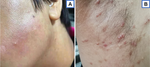
Figure 2 Histopathology examination of a skin lesion showing dense infiltrates throughout the dermis (hematoxylin-eosin, original magnification ×100; original magnification ×200) (A and B). The infiltrate was predominantly composed of neutrophils (hematoxylin-eosin staining, original magnification x 400) (C).
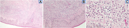
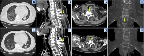
Figure 3 CT findings. 2016–3 chest CT showed multiple patchy exudations, fibrous proliferation and ground glass opacity in both lungs, bronchiectasis in the dorsal segment of the left lower lobe and pleural thickening (A). 2016–3 bone CT showed irregular bone destruction of the lower border of the C7 vertebral body, the upper border of the T1 vertebral body and its spinous process, bone defect of the anterior border of the T1 vertebral body, with surrounding abscess and the narrowing of the C7-T1 intervertebral space (arrows) (B–D). 2017–7 chest CT showed absorption of pulmonary lesions after anti-NTM therapy (E). 2017–7 bone CT shows the repair of bone destruction in C7-T1 vertebral bodies and the disappearance of the surrounding abscess after anti-NTM therapy (arrows) (F–H).