
Figures & data
Figure 1 (A) (2019–10-18) Multiple nodules, patchy high-density shadows and glass high-density shadows were seen in both lungs. (B) (2019–10-23) Multiple nodules and patchy high-density shadows were present in both lungs. (C) (2019–10-30) Nodules and patchy shadows were seen in both lungs, and bilateral pleural effusion.
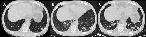
Figure 2 (A) (2019–06-27) Patchy, flocculent and ground glass-like shadows were seen in both lungs, a nodule was seen in right lung. (B) (2019–07-22) Patchy, nodular and banding shadows had significantly increased, and bilateral pleural effusion.
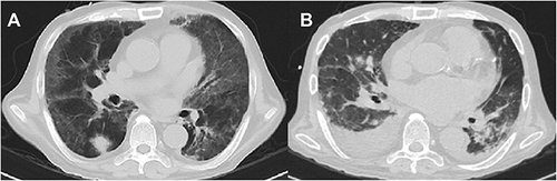
Table 1 The Clinical Characteristics, Diagnostic Methods and Other Coinfections of the 21 Patients
Table 2 The Treatment Regimens and Outcomes of the 21 Patients