Figures & data
Table 1 Summary of Cases of Infectious Keratitis After SMILE
Figure 1 A 19-year-old man with three dense round white infiltrates in the central cornea with cellular reaction in the right eye 5 days after SMILE (A). Oct-optic (cassia) examination of the right eye revealed that the central cornea was cloudy to the cap-stromal bed interface (B).
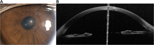
Figure 2 A 23-year-old man with a paracentral white cross infiltrate at the corneal cap–stromal bed interface 3 days after SMILE in his left eye (A). At 1 month after the surgical intervention, the cornea has only a trace of a residual peripheral scar, and the vision improved to 20/20 (B).
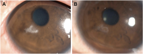
Figure 3 A 26-year-old man with a 1.5-mm melt in the central anterior corneal cap with diffuse dense infiltration at the corneal cap–stromal bed interface, and multiple keratic precipitates in the left eye 3 days after SMILE (A). Smear examination shows Gram-positive cocci (B).
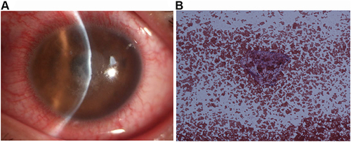
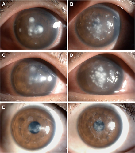
Figure 4 An 18-year-old man with several dense white satellite infiltrates of different sizes at the corneal cap–stromal bed interface, corneal edema, and endothelial immune rings 4 days after SMILE in both eyes [A(R) B(L)]. The following day, after scraping of the lesions near the original incision and interface irrigation, there is less infiltration near the incision, but increased infiltration in both eyes [C(R) D(L)]. After the incision was enlarged to about 2 quadrants, and the corneal cap was opened to fully scrape the necrotic tissue and thoroughly rinse the lesion; the infection was controlled, with significant reduction in pain and photophobia over the next 48 hours [E(R) F(L)]. At 1 year after onset, the cornea has a resolving scar, and the vision improved from hand motions to 20/32 in the right eye and 20/25 in the left eye [G(R) H(L)].
![Figure 4 An 18-year-old man with several dense white satellite infiltrates of different sizes at the corneal cap–stromal bed interface, corneal edema, and endothelial immune rings 4 days after SMILE in both eyes [A(R) B(L)]. The following day, after scraping of the lesions near the original incision and interface irrigation, there is less infiltration near the incision, but increased infiltration in both eyes [C(R) D(L)]. After the incision was enlarged to about 2 quadrants, and the corneal cap was opened to fully scrape the necrotic tissue and thoroughly rinse the lesion; the infection was controlled, with significant reduction in pain and photophobia over the next 48 hours [E(R) F(L)]. At 1 year after onset, the cornea has a resolving scar, and the vision improved from hand motions to 20/32 in the right eye and 20/25 in the left eye [G(R) H(L)].](/cms/asset/0d64cd40-2b40-4cfe-a546-838de062334b/didr_a_367328_f0004_c.jpg)
Figure 5 An 18-year-old man with several dense white infiltrates in the central cornea 4 days after SMILE in the right eye, and the infiltrates were more serious than before (A). The left eye showed multiple white infiltrates at the corneal cap–stromal bed interface (B). At 5 days after SMILE, the incision was enlarged to more than 2 quadrants, and the corneal cap was opened to fully scrape the infiltrates and rinse with antibiotics. The right eye improved 2 days later (C), but as the infiltrates worsened in the left eye (D), irrigation of the corneal cap–stromal bed interface in the left eye was repeated. At 6 months after the onset, the cornea has residual stromal scarring, and the vision improved from light perception to 20/25 in the right eye (E) and 20/32 in the left eye (F).