Figures & data
Figure 1 Gross examination of the palatine and pharyngeal tonsils, all with an unusually grey-yellowish, elastic, almost cartilaginous aspect.
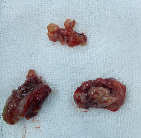
Figure 2 Clinical examination of the tongue revealing a nodular, ulcerated, infiltrating mass on the left side, extending inferiorly towards the ventral surface.
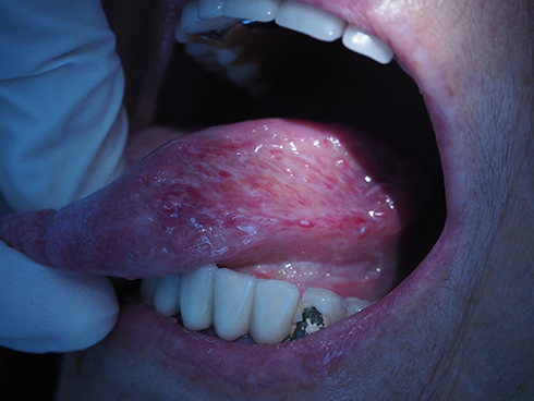
Figure 3 Microscopic examination revealing numerous caseous and non-caseous epithelioid and giant cell granulomas. (hematoxylin-eosin, 40x).
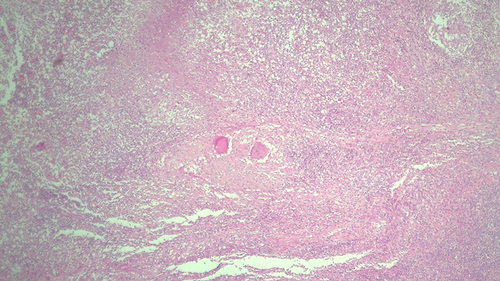
Figure 4 Ziehl-Neelsen special stain revealing acid-fast bacilli (pink rods on a blue background).
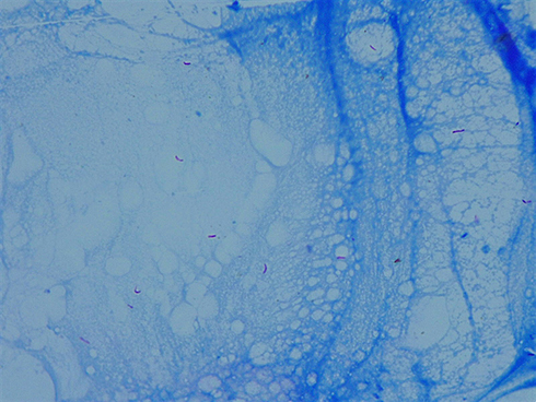
Figure 5 Clinical examination of a round, mobile, submandibular tumor.
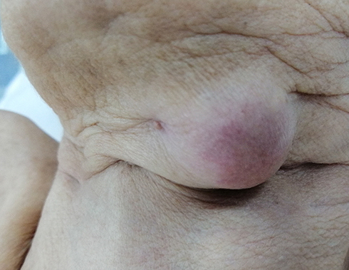
Figure 6 Microscopic examination revealing epithelioid granulomas of various shapes and sizes, necrosis, giant multinucleated cells (Langhans giant cells).
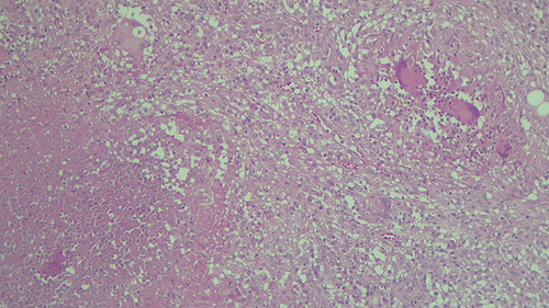
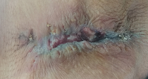
Figure 7 Defective wound healing.