Figures & data
Table 1 Minimum Inhibitory Concentration (MIC) and Antibiotics Sensitive Test (AST) of Several Antibiotics for C. Jeikeium Isolated from the CFS Culture
Table 2 Relative Abundance of Pathogenic Microorganisms in Cerebrospinal Fluid (CSF) Detected by Metagenomic Next-Generation Sequencing (mNGS)
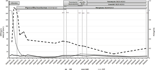
Figure 1 Brain CT plain scan results. Postoperative reexamination of intracerebral hemorrhage in the thalamus and Corona radiata areas in the right basal ganglia region revealed meningoencephalitis in the right cerebral hemisphere, and a small amount of subdural/extradural effusion in the operative area (arrow). Multiple softening foci in the brain, roughly the same as before (a-e). A contrast-enhanced brain CT on August 18 revealed a small amount of fluid within the subdural space adjacent to the right frontal lobe. An enhanced scan showed mild to moderate enhancement. There was a strip-like low-density shadow in the operative area of the right brain and a softening lesion in the right vertebral temporal lobe, which was partially connected with the adjacent ventricle. Small patches of low-density shadow were observed in bilateral basal ganglia, right midbrain, and left Corona radiate, and some edges were blurred.(A–E) No obvious abnormal enhanced shadow was observed on the enhanced scan.
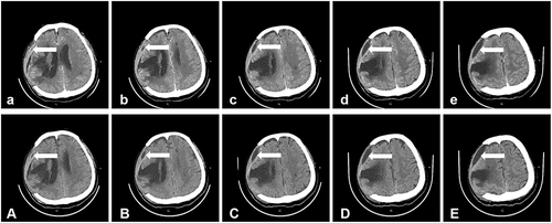
Figure 2 Brain MRI images on August 25 demonstrated post-cerebral hemorrhage in the right frontal lobe, temporal lobe, right basal ganglia, and thalamus, and the softening of the focus (a-e). The marginal enhancement of the operative area was considered to be a consequence of postoperative repair changes. The meninges indicated with the arrow was located in the right temporal region were enhanced and was considered to be inflammatory, Epidural marked curve demonstrated significant contrast enhancement following intravenous gadolinium administration (A–E) and there was slight subdural effusion in the right frontal area.
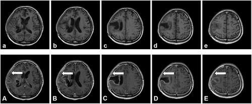
Figure 3 Clinical course of the patient with HAP and PCII in hospital.