Figures & data
Table 1 Laboratory Findings of This Case at Different Time-Points
Figure 1 X-ray radiographs at admission. Lateral (A) and anteroposterior (B) radiographs show grade I L4 spondylolisthesis and loss of height of L4–5 disc (red arrow).
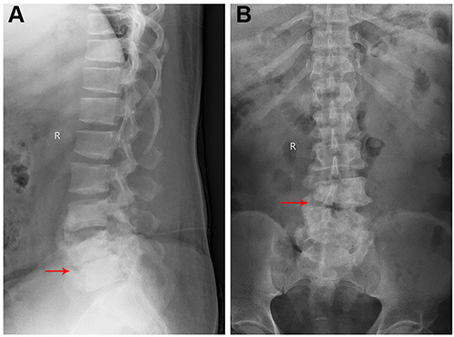
Figure 2 Fat-suppressed T2-weighted MRI images at different time-points. (A) Sagittal MRI image at admission; (B) axial MRI image of L4–5 disc space at admission; (C) sagittal MRI image at 78 days after admission; (D) axial MRI image of L4–5 disc space at 78 days after admission; (E) sagittal MRI image at re-examination; (F) axial MRI image of L4–5 disc space at re-examination. Red arrows indicate hyperintense signal in vertebral bodies and paravertebral tissue. Pink arrow indicates spinal canal abscess. Blue arrow indicates paravertebral soft tissue abscess. The damaged area was extended at 78 days after admission but relieved at re-examination.
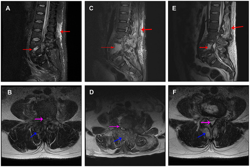
Figure 3 CT images at different time-points. (A) Sagittal CT image at admission; (B) axial CT image at admission; (C) sagittal CT image at 78 days after admission; (D) axial CT image at 78 days after admission; (E) sagittal CT image at re-examination; (F) axial CT image at re-examination. Red arrow indicates slight L3–5 vertebral body destruction with grade I L4 spondylolisthesis. Oval indicates broader and more severe L3–5 damage including grade II L4 spondylolisthesis, L4–5 disc space disappearance and L3–5 vertebral body destruction. White arrow indicates new bone formation.
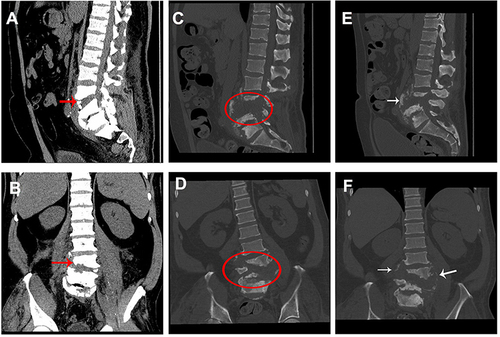
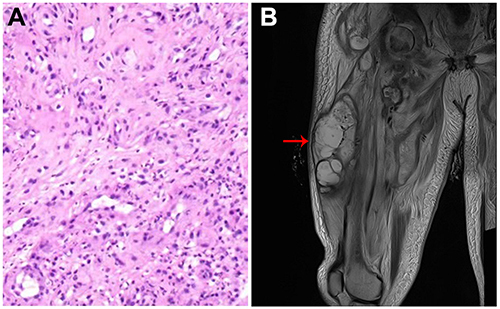
Figure 4 (A) H&E staining of L4–5 disc space tissues shows typical characteristics of acute and chronic inflammatory cell infiltration; (B) MRI image shows an obvious swelling (red arrow) in the patient’s thigh.