Figures & data
Figure 1 Head and lung CT scans of the patient after admission in our hospital. (A). Head CT showing a small amount of blood, effusion, and swelling of the surrounding soft tissue in the basal ganglia after intracerebral hemorrhage surgery. (B). Lung CT showing no obvious signs of infection.
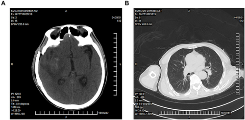
Table 1 CSF Indicators from Five Lumbar Punctures After Admission to Our Hospital
Figure 2 mNGS analysis reveals the mapping and distribution of M. hominis reads. (A) Mapping of M. hominis reads on the genome with a coverage of 3.7%. (B) (left) Total read distribution in the CSF sample, and (right) read distribution without the human host.
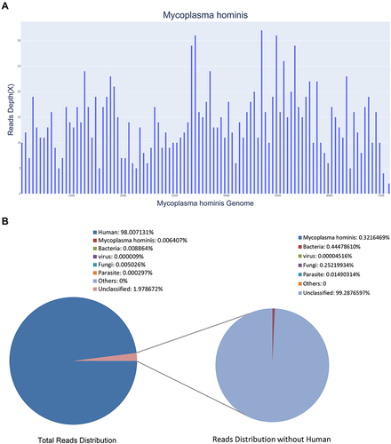
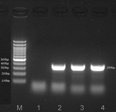
Figure 3 A 399-bp target fragment mapping to M. hominis, identified by sequence-specific PCR. Lane M: DNA ladder; Lane 1: negative control; Lane 2: annealing temperature of 58 °C; Lanes 3 and 4: annealing temperature of 60 °C.