Figures & data
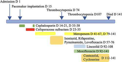
Figure 1 Chest imaging. (A) A moderate amount of liquid density shadow in the chest cavity bilaterally, mainly on the right side, with a CT value of 5 HU. Adjacent lung tissue was compressed by pleural fluid. (B) Pleural fluid was almost completely absorbed. The red arrow indicates the site of pleural fluid.
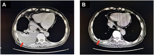
Figure 2 Antimicrobial regimens used in this patient and correlated with side effects, and interventions during inpatient days. D, patient day(s).