
Figures & data
Table 1 Four-Week Follow-Up to Assess Clinical Efficacy of Verrucae Plantaris Treatment
Figure 1 Histopathology examination before and after treatment with cantharidin cream in patients with Verruca plantaris. Patients with Verruca plantaris completed 4 weeks of treatment. (A) The histopathological examination demonstrated parakeratosis and papillomatous hyperplasia of the epidermis (hematoxylin-eosin stain, magnification 100×). (B) The histopathological examination revealed a large number of vacuolated cells in the epidermis (hematoxylin-eosin stain, magnification 400×). (C) Several neoplasms detected on the foot prior to the treatment. (D) The foot lesions were eliminated post treatment.
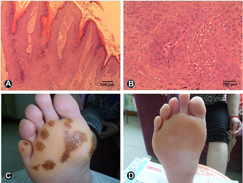
Table 2 Twelve-Week Follow-Up to Assess Clinical Efficacy of Verruca Plantaris Treatment