Figures & data
Table 1 Susceptibility Profiles of Klebsiella Pneumoniae Isolated from the Infant’s CSF, Stool Samples, and the Father’s Stool Sample
Figure 1 The string test in an isolated colony of Klebsiella pneumoniae from the CSF on an agar plate, which showed hypermucoviscosity with a > 5-mm-long viscous filament.
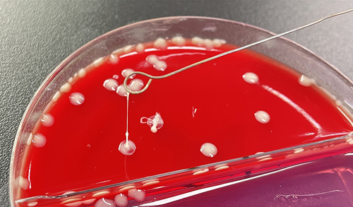
Figure 2 The virulence genes of three isolates. 1–5 represent rmpA2, iucA, peg-344, iroB, and rmpA, respectively, from the father’s stool sample, and 6–10 represent rmpA2, iucA, peg-344, iroB, and rmpA, respectively, from the infant’s stool isolate. 11–15 represent rmpA2, iucA, peg-344, iroB, and rmpA, respectively, from the infant’s CSF isolate. M represents the marker.
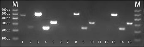
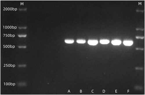
Figure 3 The capsular types of three isolates were K2. (A and B) represent the isolate from the father’s stool. (C and D) represent the isolate from the infant’s stool. (E and F) represent the isolate from the infant’s CSF. M represents the marker.