Figures & data
Table 1 Laboratory Examination of CSF in Patient with Intracranial CRPK Infection
Table 2 Bacterial Culture of CSF in Patient with Intracranial Infection
Figure 1 Brain CT after admission in patient with intracranial CRPK infection. (A) Before craniotomy on Apr 18. (B) After craniotomy on Apr 19. (C) After craniotomy on Apr 21, subdural effusion can be seen. (D) After craniotomy on Apr 24, a small amount of subdural effusion appeared. (E) After craniotomy on May 10, Subdural Effusion has developed significantly. (F) After cerebrospinal fluid leakage and intracranial infection on May 14, subcutaneous drainage tube was inserted, subdural Effusion decrease. (G) After cerebrospinal fluid leakage and intracranial infection on May 17, Subdural Effusion disappear. (H) enhanced CTbrain scanning showed no brain abscess on May 17. (I) After the treatment of anti-infective regimen on May 24. (J) After the treatment of anti-infective regimen on May 30, Ventricular Dilatation was observed.
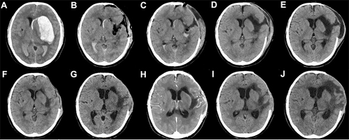
Figure 2 Location of subcutaneous drainage tube and method of compression in skull defect area. (A) Location of subcutaneous drainage tube. (B) A sport headband is used to pressurize the skull defect area.
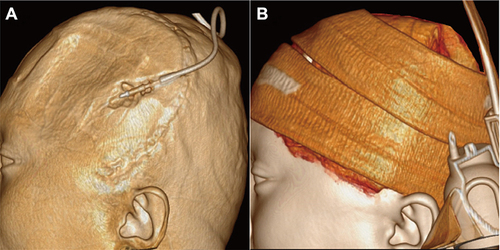
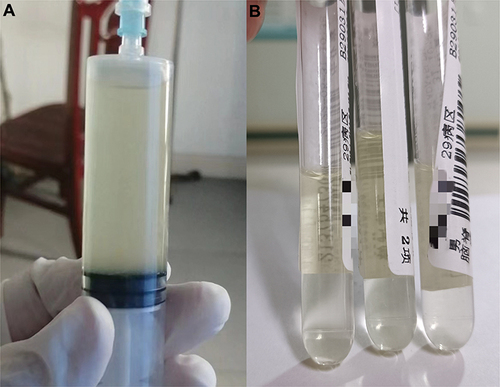
Figure 3 Changes in cerebrospinal fluid (CSF) in patient with intracranial A. baumannii infection before and after treatment. (A) CSF on May 19; (B) CSF on May 28.