Figures & data
Figure 1 The histopathological findings of the lymph node biopsy. (A) Inflammatory necrosis with granulomatous formation. Magnification, x200. Hematoxylin and eosin staining. (B) Inflammatory necrosis with granulomatous formation. Magnification, x400. Hematoxylin and eosin staining.
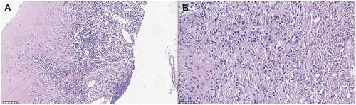
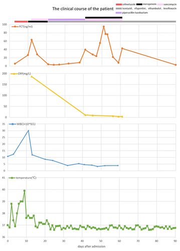
Figure 2 The clinical course of the present case. The X-axis shows the number of days after admission. The serum procalcitonin levels (Orange), C-reactive protein levels (yellow), white blood cells (blue), and temperature (green) variations were recorded. Multiple antimicrobials were given to the patient. Periods using cefmetazole, meropenem, vancomycin, piperacillin-tazobactam, and antituberculosis regimen consisting of isoniazid, rifapentine, ethambutol, and levofloxacin were marked by red, black, pink, purple and gray thick lines, respectively.