Figures & data
Figure 1 Gram-staining morphological observations of Aggregatibacter aphrophilus under the microscope. The red arrow indicates the morphology.
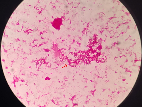
Figure 2 Images of infection caused by Aggregatibacter aphrophilus. (A) CT images of lung at admission; (B) CT images of lung 4 days after admission, the red and black arrows indicate the infection lesions and the pleural effusion on the right lung, respectively; (C) CT images of abdomen 4 days after admission, the red arrows indicate the abscess; (D) Ultrasonic images of the right psoas muscle 5 days after admission, the red arrows shows the abscess region.
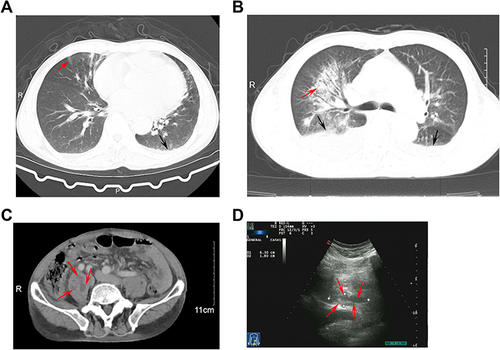
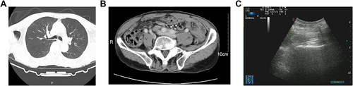
Figure 3 Images of this case after a completed treatment. (A) CT images of lung; (B) CT images of abdomen; (C), ultrasonic images of the right psoas muscle.