
Figures & data
Figure 1 Chest CT on admission showing right pleural effusion, and pneumothorax.
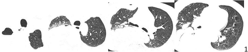
Figure 2 Against a background of necrotic fragments, a pear-shaped body with flagella was visible, containing a large dark purple long oval nucleus shaped like a mouse eye, highly suspicious for T. tenax. (indicated by arrow, Richter-Giemsa stain, ×1000).
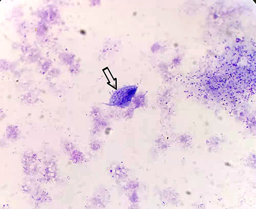
Figure 3 Chest CT at discharge showing that the right pleural effusion and pneumothorax were markedly absorbed.
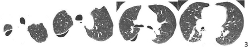
Table 1 Clinical Characteristics of the 15 Cases of Chest Infections Caused by Trichomonas