Figures & data
Table 1 Patients’ Clinical Characteristics
Table 2 CT Characteristics of the Fungal and Cancerous Nodules
Figure 1 A patient with pulmonary cryptococcosis. (A and B) axial CT images show a solid nodule located in the left upper lobe, there is a crescent-shaped collection of air and lower density in the periphery of this nodule (air crescent sign) (arrows). (B) on enhanced CT image, the nodule shows ring-like enhancement.
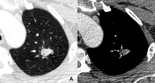
Figure 2 A patient with diabetes and pulmonary cryptococcosis. (A) axial and (B) minimum intensity projection images show a solid nodule located in the right lower lobe. There are multiple natural bronchi in it. (C) axial CT image shows that the nodule is surrounded by annular and ill-defined ground glass opacity (halo sign).
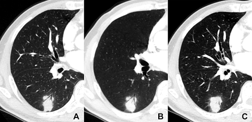
Figure 3 A patient with invasive adenocarcinoma. (A) axial and coronal (B) CT images show a solid nodule located in the right middle lobe, and there is partial and well-defined ground glass opacity (halo sign) in its periphery (arrow).
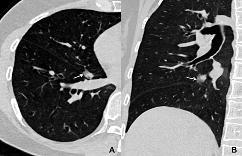
Figure 4 A patient with pulmonary cryptococcosis. (A) axial and (B) maximum intensity projection images show a solid nodule located in the right upper lobe. There are multiple scattered smaller nodules (satellite lesions, arrows) in the adjacent lung field.
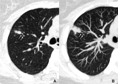
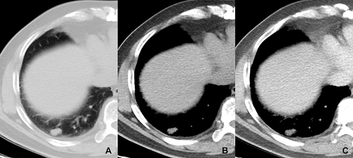
Figure 5 A patient with a history of diabetes mellitus for 15 years and pulmonary aspergillus. (A and B) axial plain CT images show an oval and well-defined solid nodule located in the right lower lobe (CT value = 14HU). On enhanced CT image (C), it has no significant enhancement (CT value = 15HU).