Figures & data
Figure 1 Cystography, renal dynamic imaging and stool imaging.
Notes: (A and B) Contrast agent leaked from the kidney to the ureter; (C) radionuclide tracing technique was used to visualize both kidneys and bladder for 10 min, 15 min and 25 min; (D) radionuclide tracing technique was also used to image stool samples.
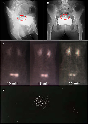
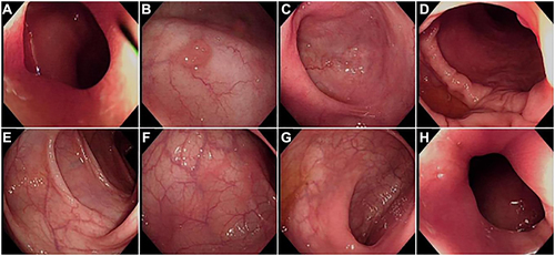
Figure 2 (A) Terminal ileum; (B) cecum; (C) ascending colon; (D) ascending colon; (E) transverse colon; (F) descending colon; (G) sgmoid colon; (H) rectum.
Notes: Multiple ulcers and erosions in the cecum, ascending colon, transverse colon, and descending colon; smooth rectal mucosa and multiple ulcers; and smooth sigmoid mucosa and scattered ulcers.
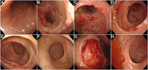
Figure 3 Pathological biopsy of the rectum, ascending colon, transverse colon, and cecum: chronic inflammation of the (rectal) mucosa.
Notes: Inflammatory exudates, necrosis and granulation tissue hyperplasia were observed in ascending colon. Transverse colon showed chronic inflammation of the mucosa, with additional inflammatory exudation, necrosis and granulation tissue hyperplasia. Cecum showed chronic inflammation of the mucosa and granulation tissue hyperplasia.
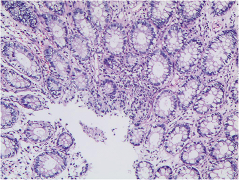
Figure 4 (A) Ileocecal orifice; (B) cecum; (C) ascending colon; (D) ascending colon; (E) transverse colon; (F) sigmoid colon; (G) rectum; (H) anal canal.
Notes: Deformation and stenosis caused by scar traction of the ileocecal valve, showing a continuous open state. White scars were observed in the cecum, ascending colon, transverse colon and descending colon, of which a few scattered polypoid hyperplasia with a diameter of about 0.2–0.5 cm was observed in the cecum and ascending colon. The mucosa of rectum and sigmoid colon was smooth, and white scars were observed in sigmoid colon, with smooth surface.