Figures & data
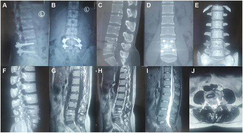
Figure 1 Radiological studies before operation. Plain radiograph showed narrowing of lumbar space 4–5 (A and B). CT showed dense pore-like destruction of the 4–5 lumbar vertebrae and intervertebral discs (C and D). Sagittal MRI showed vertebral and intervertebral disc lesions with low signal in T1, high signal in T2, and high signal in fat compression image, which was significantly enhanced after enhancement (E–H). Coronal MRI demonstrates a large, marked paravertebral abscess with unclear margins and enhancement (I and J). Axial MRI showed destruction of vertebral bodies and intervertebral discs, and significant strengthening of psoas after enhancement (K and L).
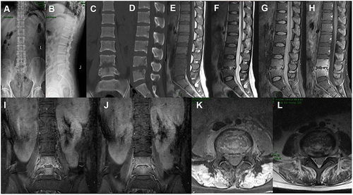
Figure 2 Intraoperatively, patients were treated with streptomycin to treat infection (A). Surgical specimens (B). Postoperative plain radiographs showed that the L4/5 vertebral space height was restored and the internal fixation was firm (C and D).
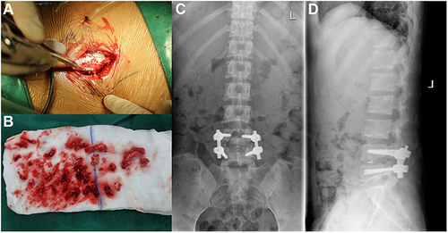
Figure 3 Brucella melitensis was detected by real-time PCR, real-time PCR showed that the DNA content of Brucella melitensis (dotted red line) increased in 30 cycles. The ordinate represents the fluorescence intensity, the abscissa represents the number of PRC cycles, the blue realization represents the baseline level, and the red realization represents the positive reference.
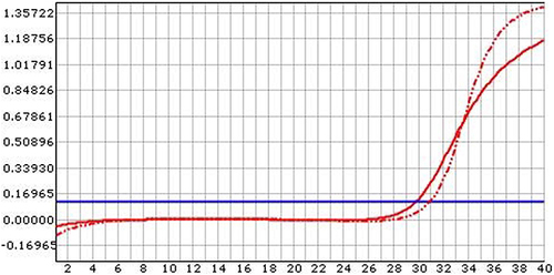
Figure 4 In postoperative pathology, Brucella (red circle) was positive for Giemsa staining (A). Mycobacterium tuberculosis was negative for acid-fast staining of the intervertebral disc of L4/5 (B).
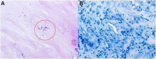
Figure 5 Radiological studies 2 years after discharge. Plain radiographs showed no abnormal changes in the fixed position of L4–L5 vertebral segments (A and B). No abnormalities on CT and MRI (C–J).