
Figures & data
Table 1 Summary of Reported Cases of Salmonella Group D Subdural Empyema
Figure 1 Neuroimaging studies of a 58-year-old male patient with Salmonella subdural empyema. (A) Axial computed tomography showing massive right convexity subdural fluid collection (white arrows) with midline shift at emergency department. (B) Axial T1-weighted magnetic resonance imaging showing thick residual subdural fluid collection (white arrows) with rim enhancement 4 days postoperatively. (C) Axial T1-weighted magnetic resonance imaging showing significantly reduced subdural empyema (white arrows) with improved midline shift before discharge. (D) Axial T1-weighted magnetic resonance imaging showing vanishing subdural empyema without midline shift after more than 5 years follow-up.
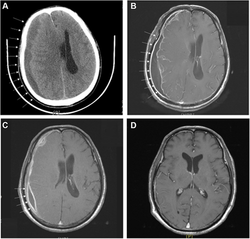
Figure 2 Intraoperative burr hole drainage gross photograph showing large amount of brown-yellow pus in subdural space.
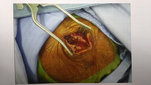
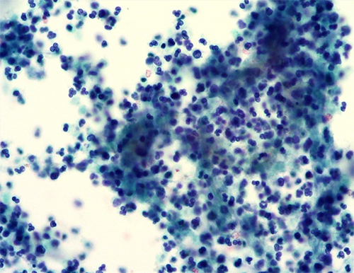
Figure 3 Histopathological photomicrograph: Papanicolaou stain, magnification ×200 showing numerous neutrophils with abundant background amorphous necrotic material, compatible with pus/abscess.
Data Sharing Statement
All clinical and other information is available in the patient’s medical record in the Division of Neurosurgery, Department of Surgery, Taipei Tzu Chi Hospital, Buddhist Tzu Chi Medical Foundation.