
Figures & data
Table 1 Antibiotic Susceptibility of Pandoraea Species from Blood and Wound Secretion Samples in Our Case
Figure 1 The first chest CT scan of the patient when she was sent to the emergency.
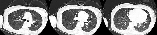
Figure 2 Bacteria colony of Pandoraea species cultured on Columbia blood plate for 48 hours.
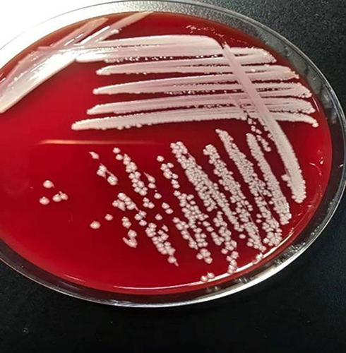
Figure 3 Wound situation of the patient before and after treatment. (A) Skin wound of the lower back; (B) skin wound of the right thigh; (C and D) Skin wound of the lower back after 2-months’ treatment.
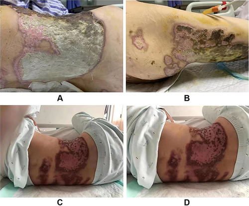
Figure 4 The treatment timeline showed clinical manifestations, significant examination results, and related antibiotic treatments of the case.
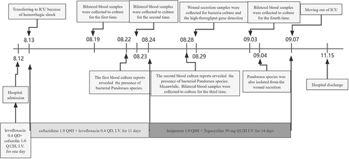
Table 2 Case Reports Infected or Colonized with Pandoraea Species in Non-CF Patients