Figures & data
Figure 1 CD86-EGFP expression in THP-1 cells via lentivirus. (A) CD86-EGFP-THP-1 cells of the OE group, (B) NC-THP-1 cells of the NC group, (C) THP-1 cells of the CON group and (D) co-culture of T. marneffei with macrophages of the CON group (the white arrows showed T. marneffei conidia) were observed under light microscopy. (E) The CD86-EGFP with green fluorescence appeared in the OE group, but no green fluorescence appeared in (F) the NC group, (G) the CON group and (H) the macrophages of the CON group after T. marneffei infection. (I) The specific CD86 protein bands of the NC and CON groups appeared at 70 kDa, and the specific protein bands of the OE group appeared at 70 kDa and 97 kDa. The band of EGFP was approximately 27 kDa, so the band at 97 kDa represented fusion protein CD86-EGFP. (J) The CD86 expression was significantly increased in the OE group in comparison with the NC group and CON group, respectively, by qRT-PCR analysis (****P < 0.0001, ns P > 0.05) (original magnification: ×400).
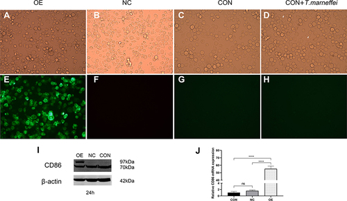
Figure 2 Live-cell imaging of co-culture of T. marneffei with CD86-EGFP-THP-1 cells by confocal microscopy. CD86-EGFP appeared with green fluorescence predominantly distributed on the cell membrane of THP-1 cells. Some of the conidia bound CD86-EGFP on the surface of the organisms. (A) T. marneffei conidia were labeled with cell membrane DIL-dye, which appeared with red fluorescence at 12 h. (B) At 24 hours, CD86-EGFP appeared as green dot-shaped particles stuck to the surface of the conidia. (C) At 24 hours, the conidia binding CD86-EGFP appeared with a body-like inclusion. (D) The conidia bind CD86-EGFP proteins, as indicated by the arrow in the top right corner. The proliferation of T. marneffei to be discovered, followed by the appearance of the fission yeasts (the arrow in the top left corner showed) at 48 h. Meanwhile, a decrease in the activity of macrophages was observed, with fluorescence intensity decreasing significantly. (E) T. marneffei conidia were cultured without macrophages. No green fluorescence was observed. (F) The image of A observed under a bright field. (G) The image of B observed under a bright field. (H) The image of C observed under a bright field. (I) The image of D observed under a bright field. (J) An image of E taken in a bright light. The white arrow showed T. marneffei conidia as the same as appeared in images of (A–E), respectively (original magnification: (A, C, D, I, F, H) ×630; (B, E, G, J) ×1000).
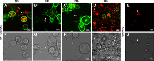
Figure 3 Live-cell imaging of CD86-EGFP-THP-1 cells infected with heat-killed T. marneffei and Cryptococcus neoformans by confocal microscopy. (A) CD86-EGFP-THP-1 cells (green) were incubated for 24 hours with DIL-labeled heat-killed T. marneffei conidia (red). It showed that there was no green fluorescence appearing in the heat-killed T. marneffei conidia. (B) For 24 hours, DIL-labeled C. neoformans (red) infected CD86-EGFP-THP-1 cells (green). It showed that C. neoformans organisms did not bind the CD86-EGFP protein. (C) The image of A observed under bright field. (D) The image of B observed under bright field (original magnification: ×630).
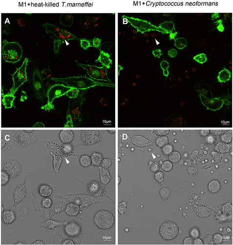
Figure 4 CD86 indirect immunofluorescence assay of THP-1 (CON group) cells co-cultured with T. marneffei conidia by confocal microscopy. (A and B) The nucleus of THP-1 cells and T. marneffei conidia were counterstained with DAPI (blue). T. marneffei conidia were highlighted by green fluorescence, which appeared as signet ring-like cells or inclusion body-like cells. (C) The image of A observed under bright field. (D) The image of B observed under bright field. The white arrow showed T. marneffei conidia as the same as appeared in images of A and B, respectively (original magnification: ×1000).
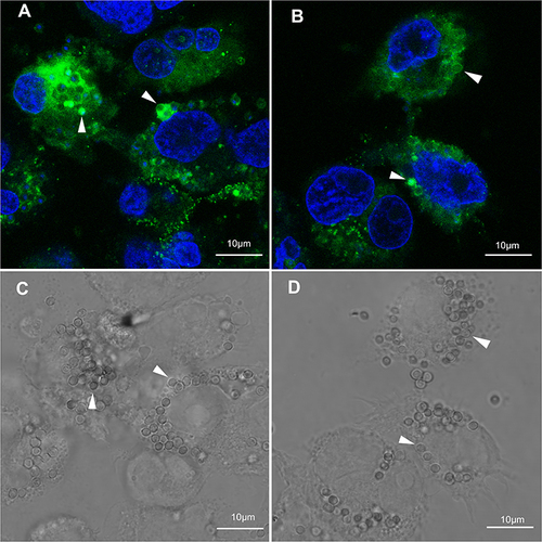
Figure 5 TCD86 expression was detected on co-culture of T. marneffei conidia with CD86-EGFP-THP-1 cells by immunoelectron microscopy. (A) Colloidal gold particles about 6 nm appeared on intracellular T. marneffei conidia of macrophage cells. (B) Colloidal gold particles of about 6 nm appeared on extracellular T. marneffei conidia of macrophage cells. (C) There were no colloidal gold particles on T. marneffei conidia cultured in medium lacking THP-1 cells (Black arrows show characteristic colloidal gold particles).
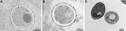
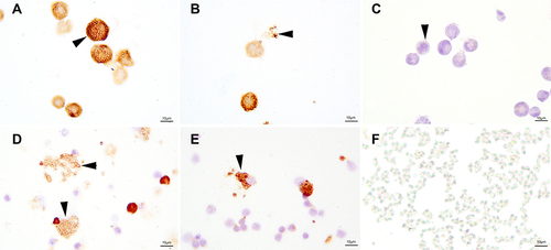
Figure 6 CD86 stain of T. marneffei conidia co-culture with THP-1 cells and PBMCs by immunohistochemistry. Mounds of T. marneffei conidia and macrophage cells had positive expression of CD86. The T.marneffei conidia with CD86 stain looked like signet rings. In (A), CD86 stained both T. marneffei conidia and THP-1 cells in the OE group. (B) In the CON group, CD86 stained both T. marneffei conidia and THP-1 cells. (C) Negative CD86 staining of T. marneffei conidia and THP-1 cells in the absence of a primary control. (D and E) T. marneffei conidia and scattered cells of PBMCs had positive expression of CD86 when the conidia were co-cultured with PBMCs. (F) Negative CD86 stain of T. marneffei conidia cultured without macrophages (original magnification: ×1000).