137
Views
1
CrossRef citations to date
0
Altmetric
CASE SERIES

Sequential Treatment by Antiviral Drugs Followed by Immunosuppressive Agents for COVID-19 Patients with Hematological Malignancy

ORCID Icon, , ORCID Icon, , & ORCID Icon
Pages 7117-7124 | Received 17 Oct 2022, Accepted 29 Nov 2022, Published online: 05 Dec 2022

Figures & data

Table 1 The Clinical Timeline of Case 1 and 2

Table 2 The Antibody Titers for SARS-CoV-2 Nucleocapsid (N) and Spike (S) Proteins in Case 1 and 2

Figure 1 Chest X-ray (A and B) and computed tomography (C and D) of the case 1 patient on admission to our hospital on day 40 (A and C) and day 52 (B and D). Interstitial shadows in X ray and Ground-glass opacities (GGOs) in CT are found in both lung fields on admission to our hospital on day 40 (A and C). These interstitial shadows and GGOs were increased 12 days later on day 52 (B and D). Both of the lung volumes were also reduced on day 52 (B), compared with on day 40 (A). Arrows indicated the abnormal shadows in chest X-ray and CT, such as interstitial shadows and GGO.

Figure 1 Chest X-ray (A and B) and computed tomography (C and D) of the case 1 patient on admission to our hospital on day 40 (A and C) and day 52 (B and D). Interstitial shadows in X ray and Ground-glass opacities (GGOs) in CT are found in both lung fields on admission to our hospital on day 40 (A and C). These interstitial shadows and GGOs were increased 12 days later on day 52 (B and D). Both of the lung volumes were also reduced on day 52 (B), compared with on day 40 (A). Arrows indicated the abnormal shadows in chest X-ray and CT, such as interstitial shadows and GGO.

Figure 2 Chest X-ray (AC) and computed tomography (DF) of the case 2 patient on day 10 (A and D), day 15 (B and E), and day 30 (C and F). GGOs are found in both lung fields on day 10 (A and D). They were worse and lesions, including the densities were increased on day 15 (B and E), but they have finally improved on day 30 (D and F). Arrows indicated the abnormal shadows in chest X-ray and CT, such as interstitial shadows and GGO.

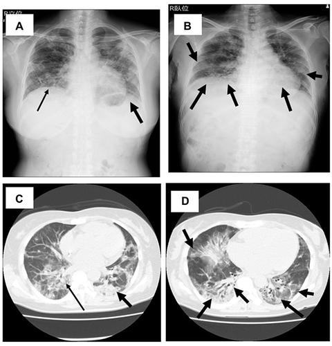
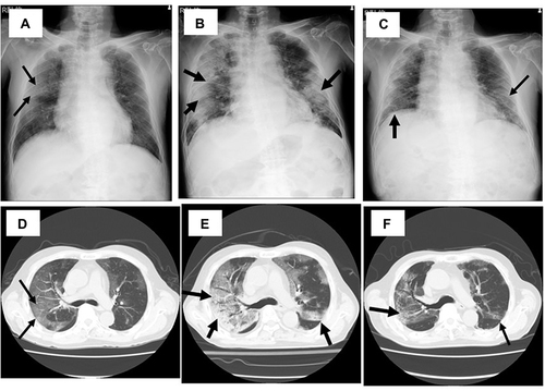
Figure 2 Chest X-ray (A–C) and computed tomography (D–F) of the case 2 patient on day 10 (A and D), day 15 (B and E), and day 30 (C and F). GGOs are found in both lung fields on day 10 (A and D). They were worse and lesions, including the densities were increased on day 15 (B and E), but they have finally improved on day 30 (D and F). Arrows indicated the abnormal shadows in chest X-ray and CT, such as interstitial shadows and GGO.