Figures & data
Table 1 Characteristics of Patients
Table 2 Characteristics of “Proven” Patients
Table 3 Characteristics of “Probable” Patients
Figure 1 The distribution of mNGS sample and infection site. (A) Distribution of mNGS samples in the “Proven” and “Probable” group. (B) The infection site in the “Proven” and “Probable” group.
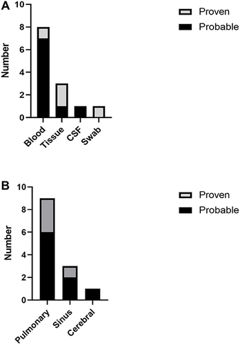
Figure 2 Imaging results of patients 1 to 4. The imaging results of patients 1–4 indicate that there may be fungal infection. (A) Chest CT indicates right pneumonia and pneumothorax. (B) Chest CT indicates bilateral pulmonary inflammation and bilateral pleural effusion. (C) Chest CT shows nodules in the anterior basal segment of the lower lobe of the left lung. (D) Imaging results showed inflammation of the right maxillary sinus and sphenoid sinus.
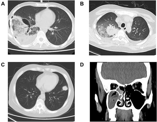
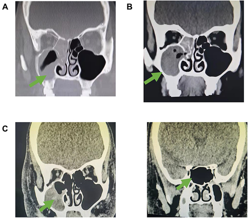
Figure 3 Imaging changes of patient 4. (A) Imaging results in D0 showed inflammation of the right maxillary sinus and sphenoid sinus, suggesting possible fungal infection. (B) The imaging results before surgical debridement (D5) indicated that the inflammation had progressed; (C) Imaging results after treatment (D16) suggest that inflammation is relieved. Arrows indicate the site of infection.