Figures & data
Figure 1 CT scan on admission (A and B), 2 months later (C and D) and 1 year later (E and F). (A and B) Axial CT images showed a thick-walled cavity in the right upper lobe containing a 4.28 cm*3.54 cm soft tissue opacity consistent with pulmonary aspergilloma and a mass of consolidation in the right lower lobe and patchy nodules shadows in the left lower lobe. (C and D) Axial CT images showed the fungal ball had been absorbed to 3.55 cm*3.00 cm and the clear left lower lobe, consolidation had been mostly absorbed. (E and F) Axial CT images showed the fungal ball had been further absorbed to a 3.09 cm*2.61 cm, and consolidation had been further absorbed.
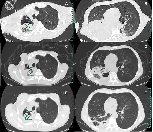
Figure 2 Macroscopic and microscopic features of Aspergillus flavus. (A) The BALF sample culturing on Columbia blood agar showed the yellow-green and cottony colonies. (B) Microscopic features including spiny conidiophores, radiant phialides on vesicles and biseriate phialides were observed under a light microscope using lactophenol cotton blue stain (LPCB, ×400).
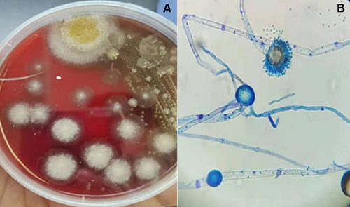
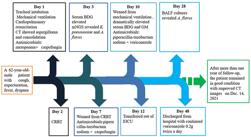
Figure 3 Treatment timeline of the reported case.