Figures & data
Figure 1 Different structures of AMPs. (A) Clavanin A, fowlicidin-1 and plantaricin A have typical α-helical conformations. (B) Gomesin, hepcidin-20 and thanatin have β-sheet conformations. (C) Termicin, phormicin and plectasin have both alpha-helix and beta-sheet conformations. (D) Drosocin, indolicidin and tritrpticin are AMPs with linear extension structures. All the structures were taken from the DBAASP v3: database.Citation112
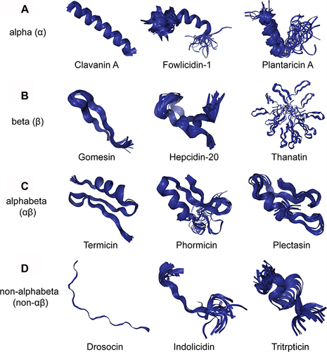
Figure 2 Three models of action for extracellular AMP activity include the carpet model, barrel stave model and toroidal pore model.Citation114
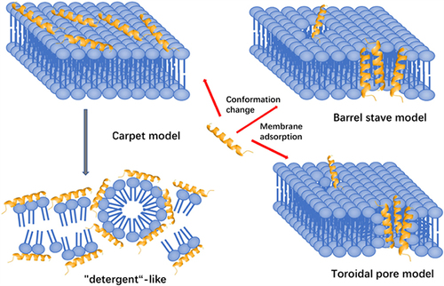
Table 1 Selection of AMPs in Clinical Phase of Development
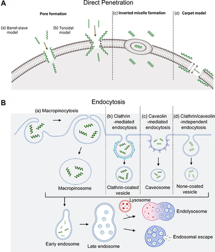
Figure 3 Different cellular uptake mechanisms of CPPs. (A) Direct translocation models including (a) the barrel-stave model; (b) the toroidal model; (c) inverted micelle formation; and (d) the carpet model.Citation82 (B) Pinocytosis models including (a) macropinocytosis; (b) clathrin-mediated endocytosis; (c) caveolin-mediated endocytosis; and (d) clathrin/caveolin-independent endocytosis. The short green curves represent CPPs.