
Figures & data
Figure 1 Chest X-ray and computed tomography images. (A) Chest images of the patient at the initial visit showing the presence of airspace consolidation in the left lung. (B) Chest images 4 months after the initial visit showing right pneumothorax (arrows). (C) Last chest image after improvement of pneumothorax.
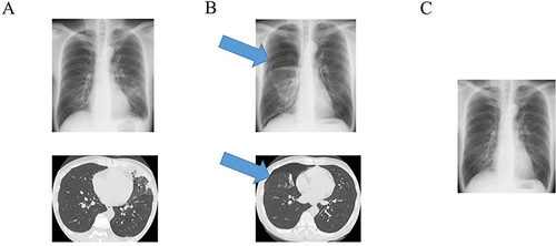
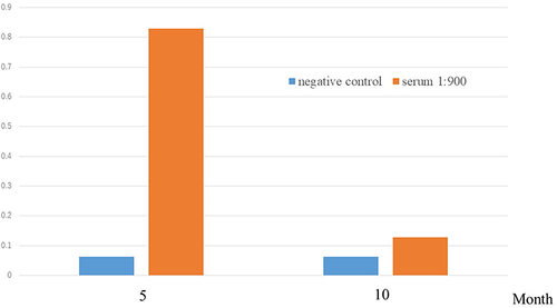
Figure 2 Antibody titer of Paragonimus westermani. Column chart showing microplate enzyme-linked immunosorbent assay analyses of Paragonimus westermani antibodies, indicating a high antibody titer at diagnosis and a decreased titer after praziquantel treatment.
Data Sharing Statement
The datasets used and/or analyzed during the current study are available from the corresponding author on reasonable request.