
Figures & data
Figure 1 Chest X-ray image showing a bilateral diffuse symmetric reticular interstitial shadow (A). A chest computed tomography scan showing bilateral asymmetric patchy mosaic appearance and ground-glass opacities in the lung subpleural peripheral regions (B and C).
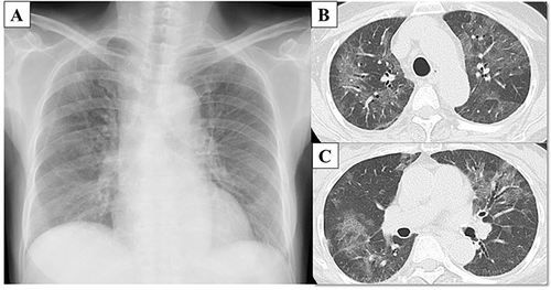
Figure 2 Clinical course of the patient in the present case.
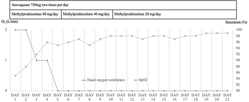
Table 1 Clinical Characteristics of the Present Patient and an Earlier Patient with Severe PCP
Data Sharing Statement
The data is available upon request.