
Figures & data
Table 1 The Level of NF-Ƙb in SMPP Group and FB Group
Figure 1 NF of FB group and SMPP group- ƙ Correlation of B (****p < 0.0001).
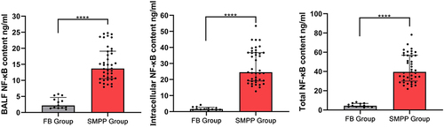
Table 2 Basic Clinical Characteristics of Children with Severe Mycoplasma Pneumoniae Pneumonia
Table 3 Correlation Analysis Between BALF MP-DNA and Clinical Characteristics
Table 4 Analysis of Correlation Between IL-6 and Clinical Characteristics
Figure 2 Correlation between clinical features (*p <0.05, red indicates positive correlation, blue indicates negative correlation).
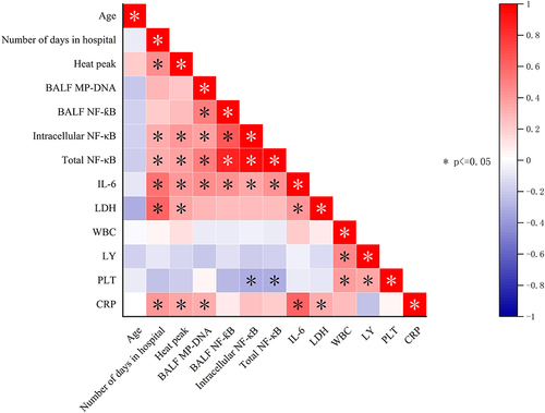
Table 5 Correlation Analysis of NF-Ƙb in BALF, NF-Ƙb in Cells, Total NF-Ƙb and Clinical Features
Table 6 NF-Ƙb Levels in Phlegm Suppository Group and No Phlegm Suppository Group
Table 7 Correlation Analysis Between BALF NF-Ƙb and Complications
Figure 3 Correlation between BALF NF-ƙB and complications (*p< 0.05).
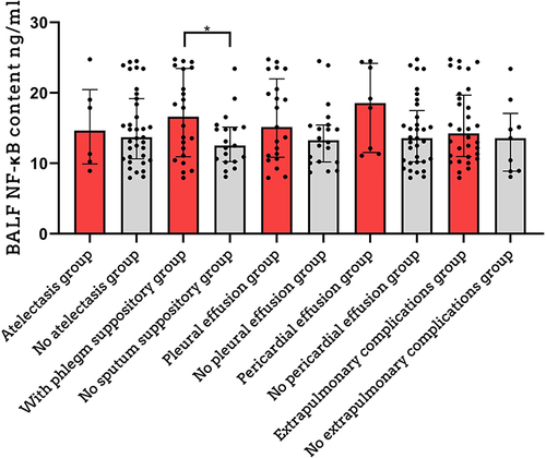
Table 8 Correlation Analysis Between Intracellular NF-Ƙb and Complications
Figure 4 Correlation between intracellular NF-ƙB and complications. (NS p > 0.05, *p < 0.05, **p < 0.01, ***p < 0.001).
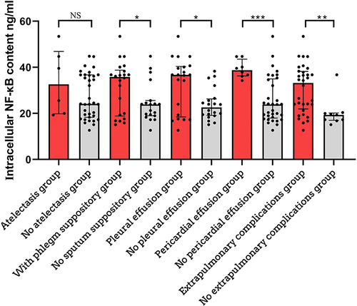
Table 9 Correlation Analysis of Total NF-Ƙb and Complications
Figure 5 Correlation between total NF-ƙB and complications (NS>0.05,*p <0.05, **p <0.01).
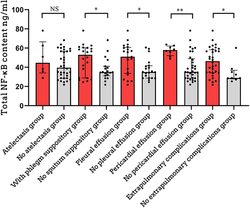
Figure 6 Chest CT changes. (A) Multilobar consolidation (high-density shadow, 3-year-old boy); (B) Consolidation of the right and left upper and lower lobes (5-year-old boy). (C) consolidation of the right upper lobe (high-density shadow, 4-year-old Boy); (D) Consolidation of the right middle lobe (hyperdense opacity, 6-year-old boy) (E) consolidation of the right lower lobe (high-density shadow, 9-year-old boy); (F) Consolidation of the left upper lobe (hyperdense opacity, 7-year-old boy). (G) Left lower lobe consolidation (high density, 5-year-old girl); (H) Atelectasis of the right upper lobe (wedge-shaped soft tissue shadow in a 9-year-old girl).
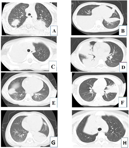
Table 10 Mucosa Changes Under Bronchoscopy
Figure 7 Mucosal changes under fiberoptic bronchoscopy. (A) 7-year-old girl with mucosal congestion and bleeding; (B) 4-year-old boy with pale mucosa; (C) 3-year-old girl with mucosal folds; (D) 11-year-old girl with mucosal bleeding points and folds; (E) 9-year-old boy with mucosal hemorrhage; (F) 5-year-old Mucous membrane erosion in a girl; (G) Sputum suppository in a 5-year-old boy; (H) Bronchial tube shape in a 4-year-old boy.
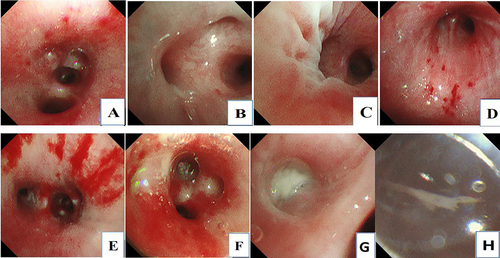
Table 11 Univariate Logistic Regression Analysis of Extrapulmonary Complications
Table 12 Multivariate Logistic Regression Analysis of Extrapulmonary Complications