Figures & data
Table 1 Burkholderia cepacia Susceptibility Results by the MIC Test
Table 2 Pathogenic Microorganisms Identified in a Subcutaneous Abscess Sample
Figure 1 Brain CT images of the patient. (A) Brain CT imaging on October 15, 2019. The head CT imaging was showed diffuse edema of brain tissue, gray matter and white matter structure blurred. (B) Brain CT on September 14, 2022. The brain CT imaging was showed the right temporo-parietal cerebral hemorrhage and occipital defect with subcutaneous abscess.
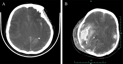
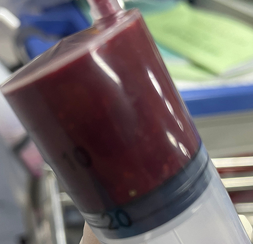
Figure 2 The sampling of bloody pus.