
Figures & data
Figure 1 Imaging changes documented the alleviation of brain lesions during the timeline of treatment (a) The initial image of brain MRI at external healthcare center, and the section that indicated by red arrow represented intracranial space-occupying lesions. (b) The image of brain MRI before surgery in our hospital, the region that indicated by red arrows suggested further exacerbation of brain lesion. (c) The image of brain MRI in early postoperative, the region that indicated by red arrow documented the removal of cerebral abscess and existence of perilesional edema. (d) The brain CT scan of day 25 after surgery, documented recovered well of brain lesions without a relapse.
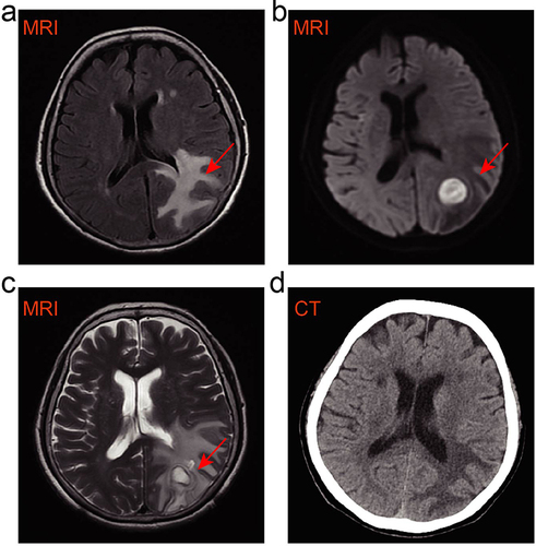
Table 1 Identification of Nocardia Genus in Cerebral Abscess Using mNGS
Figure 2 Microbiology detection of N. gipuzkoensis. (a) The traditional culture of cerebral abscess with the pale yellow, rough, and dry colonies on Columbia Blood Agar medium. (b) The characteristic of weakly acid-fast was identified by acid-fast staining.
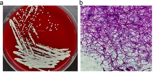
Table 2 Antimicrobial Drug Susceptibility Testing of Nocardia Gipuzkoensis
Table 3 Summarizes Some Data from the Case Literature of Cerebral Abscess Infected by Nocardia spp. in Patients