Figures & data
Figure 1 MRI and CT findings of the patient. (A–C) The MRI examination showed the destruction of vertebral at thoracic 4–7 (mainly 5–6) on October 11, 2022. (D–F) The CT examinations revealed the destruction of the vertebral bodies of thoracic 5–6 and cervical 6–7 at different times. (D–F) represent the CTs on October 11, October 21 and November 26, respectively. The destruction of the thoracic 5–6 was well improved when compared to the CT on admission. The red rectangles indicate the region of thoracic 5–6.
Abbreviations: T2WI, T2 weighted imaging; FS-T2WI, fat saturation-T2 weighted imaging; T1WI, T1 weighted imaging; Oct., October; Nov., November.
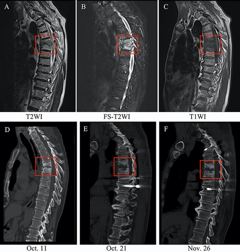
Figure 2 Rose-Bengal plate test for brucellosis. (A) Negative for saline control; (B) Positive for agglutination of the patient’s serum.
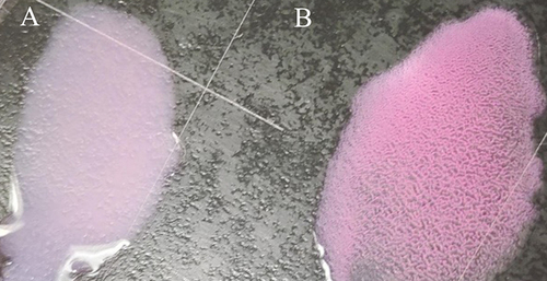
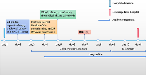
Figure 3 Timeline of the patient’s hospitalization and discharge.