
Figures & data
Table 1 Demographics and Clinical Characteristics of Patients with C. psittaci Infection
Figure 1 Cumulative hospital admissions of psittacosis per month.
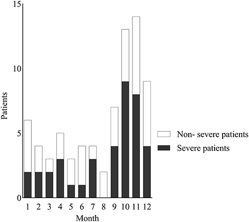
Table 2 Laboratory Findings and Chest CT Findings Upon Hospital Admission with C. psittaci Infection
Figure 2 Chest imaging of a 67-yr-old man with C. psittaci infection show multiple consolidations in the right upper and lower lobe, with air bronchogram in the upper lobe.
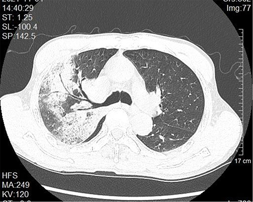
Figure 3 Chest imaging of a 78-yr-old man with C. psittaci infection before and after treatment. (A) Pre-treatment computed tomography (CT) scan showing bilateral large consolidation with air bronchogram in right lung and bilateral pleural effusion. (B) Follow-up CT scan after combination therapy for 20 days showing the consolidation and pleural effusion disappeared.
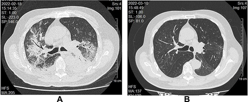
Table 3 Complications, Treatments and Outcomes of Patients with C. Psittaci Infection
Data Sharing Statement
The datasets generated and analyzed during the current study are available from the corresponding author upon reasonable request.