
Figures & data
Figure 1 (a) Chest CT showed pulmonary inflammation; (b) Brain CT showed ventriculomegaly on admission.
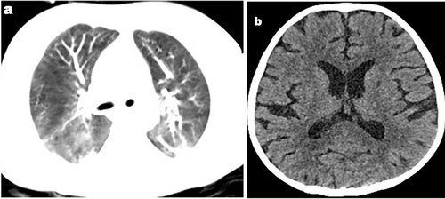
Figure 2 (a) The variation of the patient’s temperature; (b) The variation of CRP; (c) The variation of leukocytes; (d) The variation of thrombocytes.
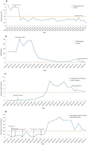
Table 1 Susceptibility Results for P. Aeruginosa in Blood Samples
Table 2 Susceptibility Results for P. Aeruginosa in Sputum Sample
Figure 3 (a) Chest CT showed pulmonary inflammation relieved, but cavitary lesions formed (black arrows); (b) Brain MRI of the brain revealed ventriculomegaly, along with abnormal signal intensities observed in the bilateral precornu, the left frontal lobe, and the triangular region of white matter (black arrows).
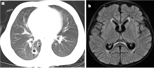
Figure 4 The timeline of the treatment and follow-up.
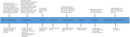
Table 3 Neonatal and Pediatric Cases Receiving Intrathecally or Intraventricularly of Polymyxin B
Data Sharing Statement
Data will be provided by the corresponding author upon reasonable request.