Figures & data
Table 1 Material Characteristics of the Implant
Table 2 Complete Set of Surgical Instruments for Vertebroplasty
Figure 1 The VAS scoring criteria for low-back pain.
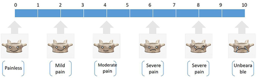
Figure 2 Cobb angle method for waist measurement.
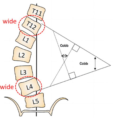
Figure 3 Flow chart of test grouping.
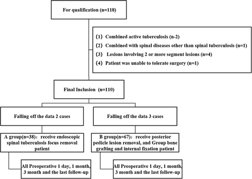
Table 3 Basic Preoperative Data of the Two Groups
Table 4 Postlumbar VAS and ODI Scores in Both Groups
Table 5 Test Tested at 1 Day and Last Follow-Up in the Two Groups
Table 6 Comparison of Postoperative Complications Between the Two Groups
Table 7 Comparison of Transfusion Rates and Transfusion Volume Between Two Groups
Table 8 Comparison of Secondary Indicators Between the Two Patient Groups
Figure 4 Box plots of comparative secondary indicators in the two patient groups.
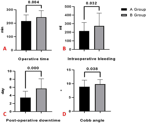
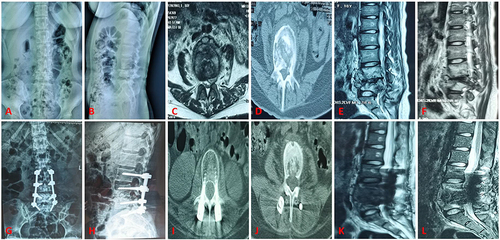
Figure 5 Group A is a typical case ①.
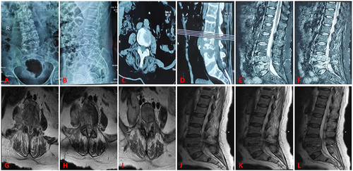
Figure 6 Group A is a typical case ②.
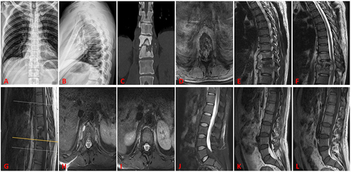
Figure 7 Group B is a typical case ①.
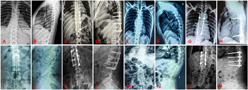
Figure 8 Group B is a typical case ②.