
Figures & data
Figure 1 (A) Obvious conjunctival congestion, with white opacity on the temporal side of the cornea and visible brown foreign body in his right eye; (B) Confocal microscopy revealed abundant hyphae structure at the lesion of the right cornea; (C) Calcofluor white staining of corneal scrapings showed abundant septate hyphae.
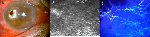
Figure 2 (A) The colonies on PDA and SDA after 7 days incubated at 25°C and 35°C; (B and C) Micromorphology examination showed hyphae hyaline and chlamydospores. Chlamydospores produced laterally or intercalary on hyphae, chlamydospores produced laterally or intercalary on hyphae, single-cell, thick-walled, brown, globose to subglobose.
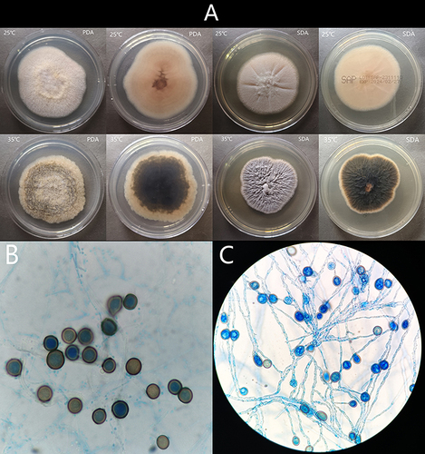
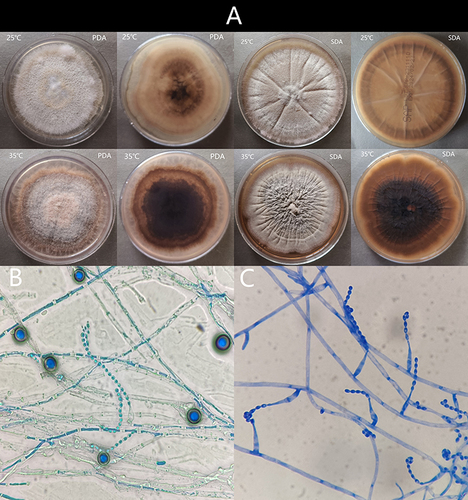
Figure 3 (A) The colonies on PDA and SDA after 14 days incubated at 25°C and 35°C. (B) Micromorphology examination showed two types of conidia: chlamydospores and acremonium-like conidiophores; (C) Acremonium-like conidiophores, which were small (2.5–3 µm × 1.2–1.8 µm), hyaline, aseptate, smooth, obovoid, with slightly truncate ends, arranged in long divergent chains on the apex of the flask shaped conidiogenous cells (phialides).
Data Sharing Statement
The datasets used and analysed during the current study available from the corresponding author on reasonable request.