Figures & data
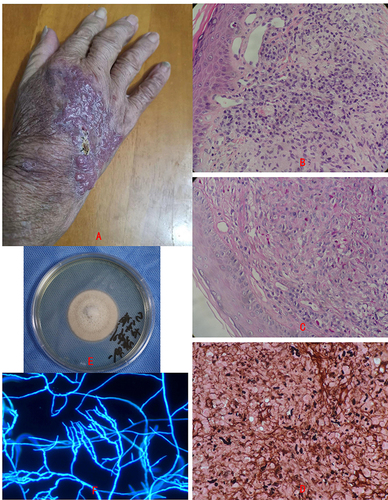
Figure 1 Clinical aspect and biopsy of phaeohyphomycosis patient and the culture of Paraconiothyrium cyclothyrioides. (A)The clinical aspect of the patient, is crusted, ulcerated plaque on the right opisthenar and several macular papules and nodules on the erythematous plaque. (B–D) Biopsy specimens revealed pseudoepitheliomatous hyperplasia and suppurative granulomas, and fungal elements were seen invading the dermis by PAS and GMS (×400). (E) Culture of the tissue specimens yielded a one colony type mold. (F) Microscopic morphology showed septate, bamboo-like hyphae, and no spores were found (×400).