Figures & data
Figure 1 (A and B) Bilateral lateral rectus palsy.

Figure 2 Clinical course of the patient.
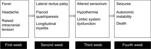
Figure 3 MRI showing thalamic hyperintensities (arrows). T2 weighted image (A); diffusion weighted image (B).
Abbreviation: MRI, magnetic resonance imaging.
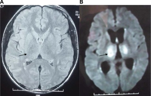
Figure 4 Longitudinal myelitis extending caudally upto L1 and rostrally upto pons. T2 weighted image (A); T1 weighted image (B). The white arrows show white matter hyperintensities.
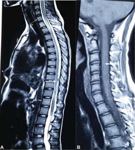
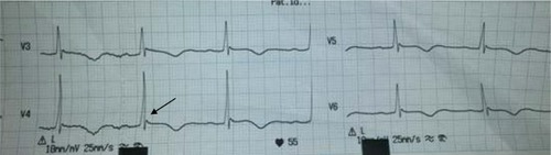
Figure 5 ECG showing bradycardia, prolonged QT interval and Osborn Wave (arrow).
Abbreviation: ECG, electrocardiography.