Figures & data
Table 1 Clinical presentation of Nasopharyngeal bursitis
Figure 1 Thirty-degree rigid endoscopic appearance of nasopharyngeal bursitis. A) Crust type. Note the characteristic midline anatomic site with cicatricial streaks around the bursa. B) Cystic type. Photos are representative of three patients of each type showing very similar appearance. C) One-year postoperative endoscopic view of the crust type.
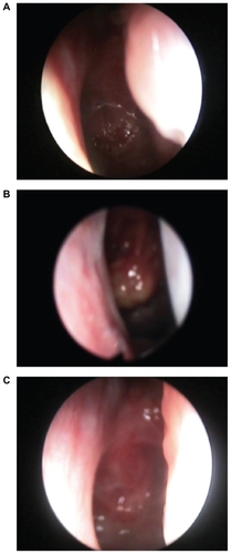
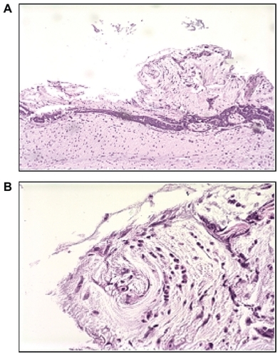
Figure 2 A) Low and B) high power fields showing histologic appearance of crust type bursitis showing reactive lymphoid mucosa with necrotic tissue.