Figures & data
Figure 1 Initial CT-scan showing abscess with air–fluid level abutting left psoas muscle, indicated by red arrow.
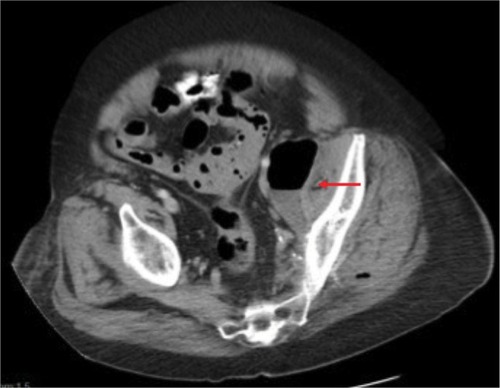
Figure 2 Repeat CT-scan showing abscesses in the left gluteal muscles, indicated by red arrows.
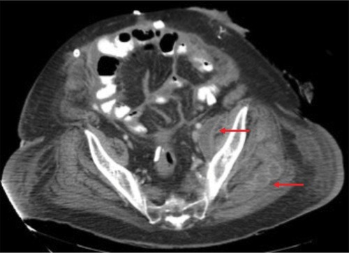
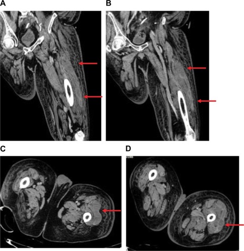
Figure 3 (A, B) CT-scan of left lower extremity, coronal sections showing abscess spanning the length of the vastus lateralis muscle. (C, D) Axial slices showing extent of abscesses. Red arrows indicate abscesses in vastus lateralis muscle.