Figures & data
Figure 1 Abdomen CT with intravenous and oral constrast demonstrating swelling of appendix, thickened wall of cecum and perifocal fat stranding (A). The fluid-contained hernia sac was seen lateral and inferior to the pubic tubercle, with circumferential fat stranding (B).
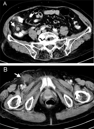
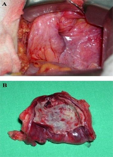
Figure 2 Intraoperative findings showed contricted hernia neck of the femoral hernia without herniation of abdominal viscera (A), and acute phlegmonous inflammation of the incarcerated hernia sac (B).