Figures & data
Figure 1 Mid-sagittal T1-weighted image of the brain shows a well-defined, rounded lesion at the roof of posterior third ventricle exhibiting low to intermediate signal intensity relative to the gray matter, with layering at the dependent portion.
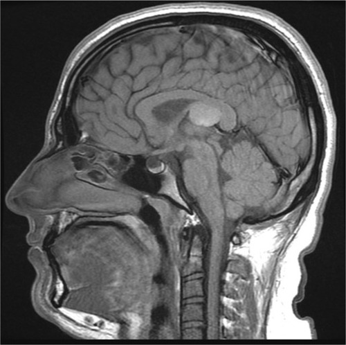
Figure 2 Axial T2-weighted (A) and fluid-attenuated inversion recovery (FLAIR) (B) images of the brain showing a well-defined, rounded lesion at the posterior part of the third ventricle.
Notes: The lesion exhibited homogenous, low to intermediate signal intensity and indented the wall of the lateral ventricle. The foramina of Monro were patent, with no signs of hydrocephalus.
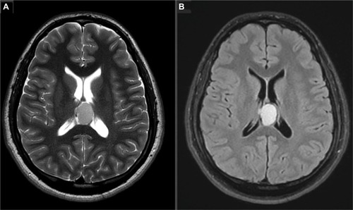
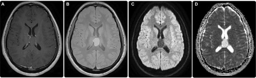
Figure 3 Post-contrast images showing no enhancement (A). Gradient images indicating no blooming artifacts to suggest hemorrhage or calcification (B). No diffusion restriction was elicited in diffusion-weighted imaging (DWI) (C)/apparent diffusion coefficient (ADC) (D) sequences.