Figures & data
Figure 1 Colonoscopy findings including Indigo carmine spraying (A, ascending colon; B, cecum; C, rectum).
Notes: (A) Mass of the submucosal-like tumor. (B and C) Isolated small polypoid tumors. An abnormal microvascular tree was noted on the surface of these lesions.
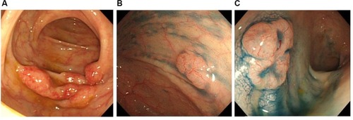
Figure 2 Histopathology findings (A, hematoxylin–eosin staining; B, CD20 immunostaining; C, BCL2 immunostaining, ×100).
Notes: (A) Small- to medium-sized lymphoid cells with mild atypia proliferation and follicular patterns were observed. (B and C) The tumor cells stained strongly positive for CD20 and BCL2.
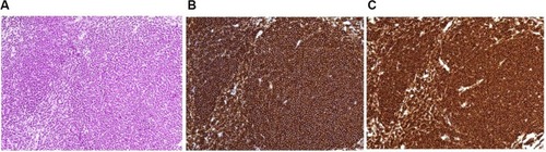
Figure 3 IgH/BCL2 rearrangement by FISH analysis.
Notes:
IgH probe for 14q32 (green signals) and BCL2 probe for 18q21 (orange signals) and arrows (yellow signals) indicate IgH/BCL2 fusion. We detected IgH/BCL2 fusion in 95.2% of tumor cells.
Abbreviation: FISH, fluorescence in situ hybridization.
Abbreviation: FISH, fluorescence in situ hybridization.
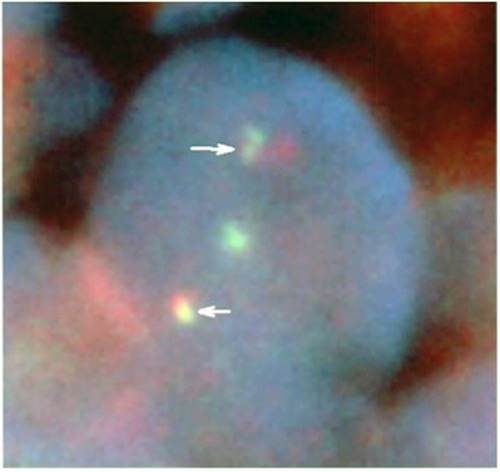
Figure 4 18F-FDG-PET/CT finding.
Notes: In the ascending colon, abnormal uptake that was consistent with malignant lymphoma was observed. SUVmax was 4.09.
Abbreviations: CT, computed tomography; 18F-FDG-PET, [fluorine-18]-fluorodeoxy-glucose-positron emission tomography; SUVmax, maximum standardized uptake value.
Abbreviations: CT, computed tomography; 18F-FDG-PET, [fluorine-18]-fluorodeoxy-glucose-positron emission tomography; SUVmax, maximum standardized uptake value.
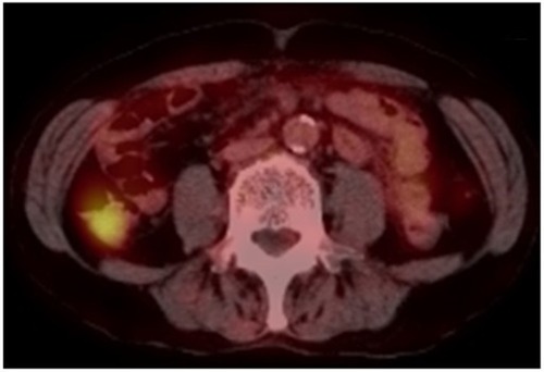
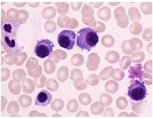
Figure 5 Bone marrow aspiration smear (May–Giemsa staining, ×1,000). Note: The frequency of plasma cells was 30% of all nucleated cells.