Figures & data
Figure 1 Left: Preoperative abdominal CT. Solid hepatic lesion at segments VI and VII (arrows) and calcified lesion at the tail of the pancreas (circle). Right: Postoperative abdominal CT. Subcapsular hepatic fluid collection without recurrence of the HCC (circle).
Abbreviations: CT, computed tomography; HCC, hepatocellular carcinoma.
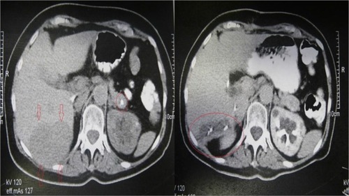
Figure 2 (A) Scar lesion from the pancreas (H&E 100X). (B) The tumor cells are arranged in abnormal and thick trabeculae (H&E 100X). (C) The neoplastic cells are positive for HepPar1 (Immunostain 200X). (D) The neoplastic cells are positive for AFP (Immunostain 200X).
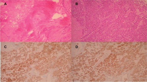
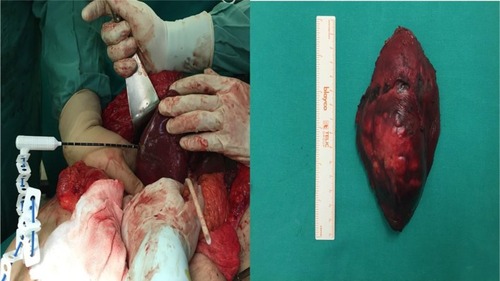
Figure 3 Left: Intraoperative photo. Microwave tissue coagulator marking the margins for hepatic segmentectomy. Right: Gross specimen photo. Hepatic segments VI and VII containing the HCC.
Abbreviation: HCC, hepatocellular carcinoma.