Figures & data
Table 1 Characteristics and pulmonary function test results of SM-injured patient and control group
Table 2 Sequence and features of PCR
Figure 1 MT-1A gene expression in SM-injured patients and in unexposed control cases. Gene expressions were measured by semiquantitative reverse transcriptase polymerase chain reaction, and were upregulated in SM-injured patients (lanes 3–12). Normal samples (lanes 1 and 2), SM-injured patients samples (lanes 3–13).
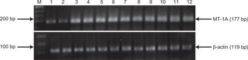
Table 3 Increases in MT1A expression in sulfur mustard-exposed patients in comparison with control group
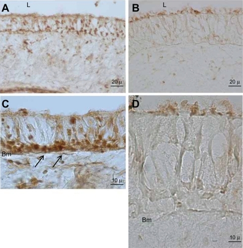
Figure 2 Immunohistochemical staining for MT-1A protein in bronchial epithelium. Left side micrographs reveal the expression of MT-1A in the basal cell layer of control group airway epithelium (arrow, A and C). Right side micrographs show MT-1A expression in SM-injured airway epithelium (B and D). Note the immunoreactivity of MT-A1 in the control group is higher and wider than in the SM-injured samples. In the high and same magnification the epithelium layer thickness in SM-injured is much thicker than control group (C and D).