Figures & data
Table 1 Classification of EB
Table 2 Differential diagnosis of EB
Table 3 Summary of important recommendations in the guidelines for EB pain assessment
Table 4 Complications associated with EB and their management
Figure 1 Appearance of lesions on the patient’s back on presentation.
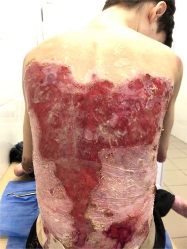
Figure 2 Appearance of lesions on the patient’s chest and abdomen on presentation.
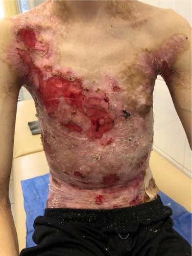
Figure 3 Appearance of lesions on the patient’s chest and abdomen on presentation.
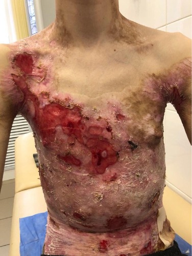
Figure 4 Appearance of deformities on the patient’s hands on presentation.
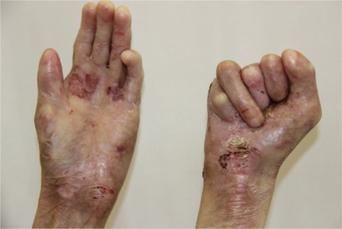
Figure 5 Appearance of lesions on the patient’s elbow on presentation.
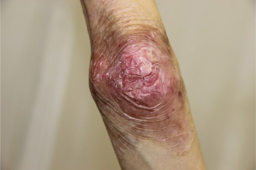
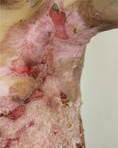
Figure 6 Appearance of lesions on the patient’s lateral abdomen on presentation.
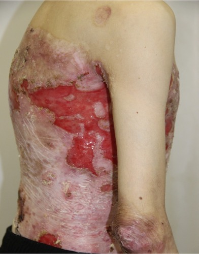
Figure 7 Result of the patient’s skin biopsy showing fixation of IgG in the basement membrane of the epidermis, on the roof the subepidermal blisters, with minor migration in the intercellular space of the epidermis and moderate in the papillary layer of the dermis.
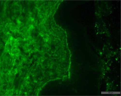
Figure 8 Result of the patient’s skin biopsy showing fixation in the basal membrane of the epidermis, and slightly in the dermis, with permeation of some keratinocytes of the C3 component of complement.
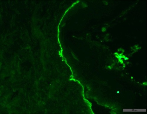
Figure 9 Appearance of lesions on the patients chest and abdomen on day 1 after discharge.
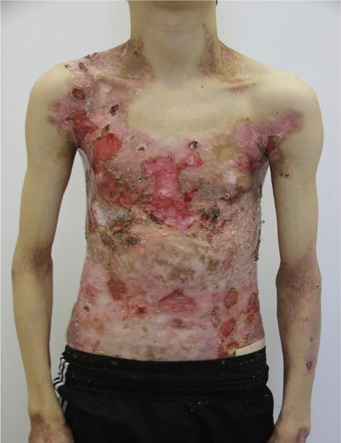
Figure 10 Appearance of lesions on the patients axilla on day 1 after discharge.