Figures & data
Figure 1 A transabdominal ultrasound scan showing the mass (red arrow) and the uterus (black arrow), bladder (blue arrow).

Figure 2 Pelvi-abdominal MRI showing a large, well-defined, oval mass measuring 12 × 8 cm in dimensions overlapping the right side of the pelvic cavity (red arrow). The mass appears to be connected to the uterus (blue arrow). A subserosal leiomyoma is suggested. However, a desmoid tumor needs to be excluded.
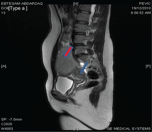
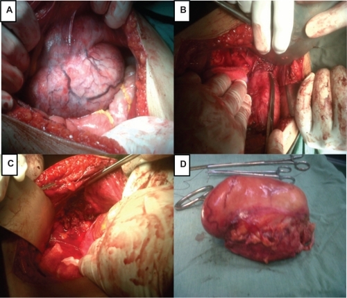
Figure 3 A) A picture showing the tumor in the inner aspect of the abdominal wall. B) The picture shows the margin of the sharp dissection. C) The tumor has been completely removed with a safe margin. D) The whole tumor after removal.