Figures & data
Figure 1 Cardiomegaly and dilatation of right and left lung hilum. Increased vascular shadowing in both sides. Costophrenic regions free of pleural effusion.
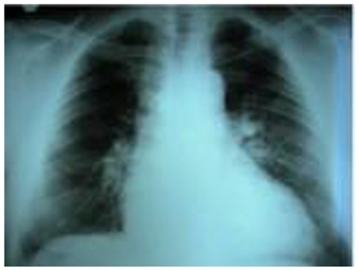
Figure 2 High probability for pulmonary embolism ventilation/perfusion scan demonstrating normal ventilation and multiple mismatched segmental and larger defects more in the right lung. The rate of perfusion for the right and the left lung is 27.4% and 72.4%, respectively.
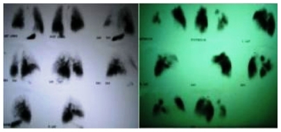
Figure 3 Computed tomographic angiogram demonstrating findings of chronic pulmonary embolism and showing occlusive thrombus in the right pulmonary artery and its branches for the right lower lobe. Embolus within the left main pulmonary artery extending into the lobar branches. Small pericardial effusion, enlarged right ventricle, and congestion of inferior vena cava and hepatic veins.
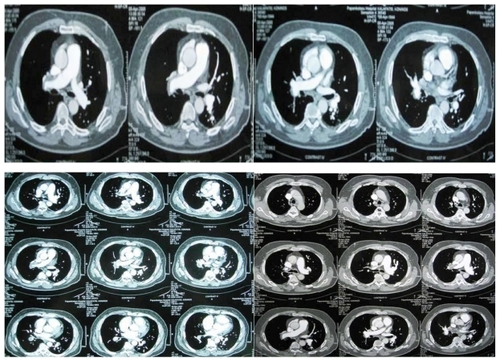
Table 1 Patient’s clinical history