Figures & data
Table 1 Laboratory Tests Related to Bone and Mineral Metabolism
Table 2 Changes in the Results of DEXA
Figure 1 MRI T2 images (A and B) of the right wrist showed Barton fracture with marrow edema. MRI T2 images (C) showed no fractures in lumbar vertebrae.
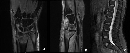
Figure 2 Preoperative X-ray films (A and B) and three-dimensional reconstructive CT images (C and D) of right wrist. Postoperative X-ray films of right wrist (E and F).
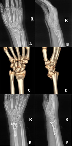
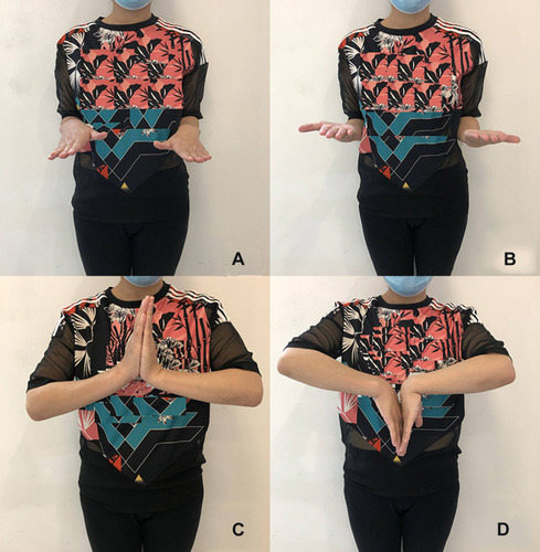
Figure 3 Follow-up after 3 months of surgery of the patient. The pronation and supination of forearm (A and B). The flexion and extension functions of wrist (C and D).