Figures & data
Figure 1 Supine abdominal radiograph showed a large gas-filled dilated bowel loop in the left mid and lower quadrants. The intestinal pneumatosis (arrow) can also be seen in the upper and right lower abdomen.
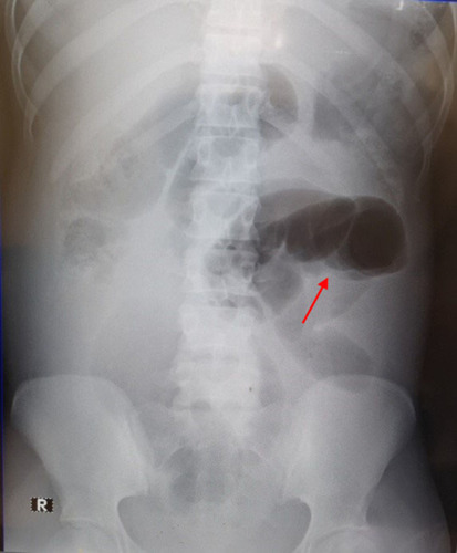
Figure 2 Intraoperative photographs show (A) gangrenous sigmoid colon, (B) gangrenous ileum loop and (C) cecum. Arrow: terminal ileum.
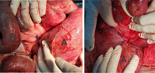
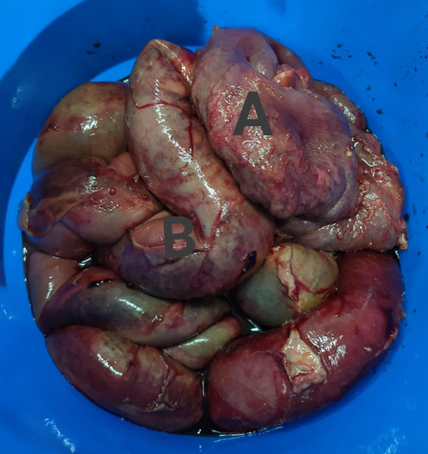
Figure 3 Postoperative photograph shows the surgical specimen (A: gangrenous sigmoid colon; B: gangrenous ileum loop).