Figures & data
Table 1 TdT Immunoreactivity in GCTs and Sex Cord-Stromal Tumors
Table 2 TdT Immunoreactivity in GCTs in the Most Recent 3 Years
Table 3 Comparation of TdT Staining to That of PLAP, OCT4, SALL4, CD117, D2-40 in GCTs (Chi-Square/Fisher Exact Test)
Figure 1 Immunohistochemical staining of TdT in seminomas. Seminomas are characterized by diffuse sheets or nests of tumor cells separated with lymphocytic infiltration; tumor cells have relatively uniform and large nuclei, prominent nucleoli, and abundant clear or eosinophilic cytoplasm (A). Tumor cells were diffusely and strongly positive for TdT (B) and confirmed by OCT4 (C). Old archived tissues may lose TdT antigen to varying degrees and therefore give a misinterpretation of “negative” staining for TdT (D, highlighted between the black arrowheads) (amplification: 10×20, bar=50μm).
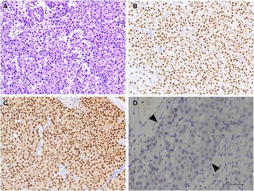
Figure 2 Immunohistochemical staining in GCNIS. GCNIS features large atypical tumor cells located (arrow) on the periphery of seminiferous tubules with prominent nucleoli and abundant clear cytoplasm (A). The tumor cells were strongly positive for TdT (B). These GCNIS cells are confirmed by OCT4 (C) (amplification: 10×20, bar=50μm).
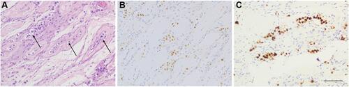
Figure 3 Immunohistochemical staining in dysgerminoma. Dysgerminoma presents with sheets of relatively uniform tumor cells with intervening thin fibrous septa and small lymphocytes (A). The tumor cells were diffusely and strongly positive for TdT (B) and confirmed by OCT4 (C) (amplification: 10×10, bar=50μm).
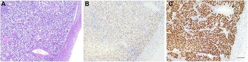
Figure 4 Immunohistochemical staining in EC. ECs demonstrate heterogeneous growth patterns, including solid, complex glandular, or papillary figurations (A). Positivity for TdT in EC (B). The tumors were confirmed by OCT4 (C) (amplification: 10×20, bar=50μm).
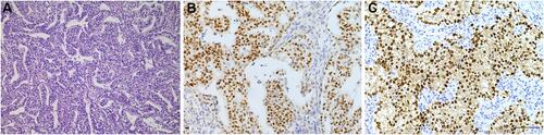
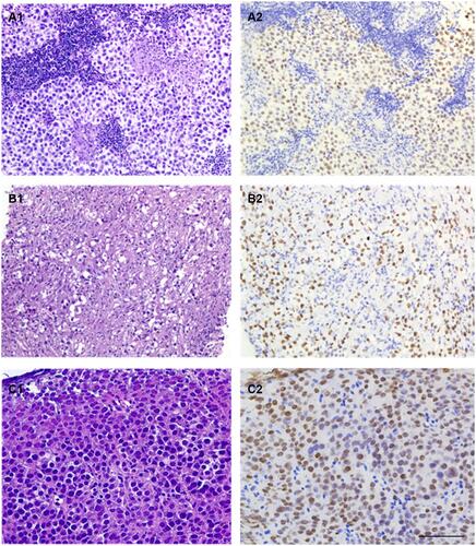
Figure 5 Extragonadal germinomas involving the sellar (A1), mediastinal (B1), and right cervical (C1) regions were obtained through core biopsies. The morphologic features are identical to the testicular or ovarian counterpart; crush artifact and fibrous changes (B1) are not uncommon. All of these tumors express TdT (A2–C2) (amplification: 10×20, bar=50μm).